Pneumothorax: Collapsed Lung Symptoms and Emergency Care Guide
 Mar, 27 2026
Collapse isn't just a term for buildings; it describes exactly what happens when air escapes your lung tissue.
Understanding the Sudden Collapse
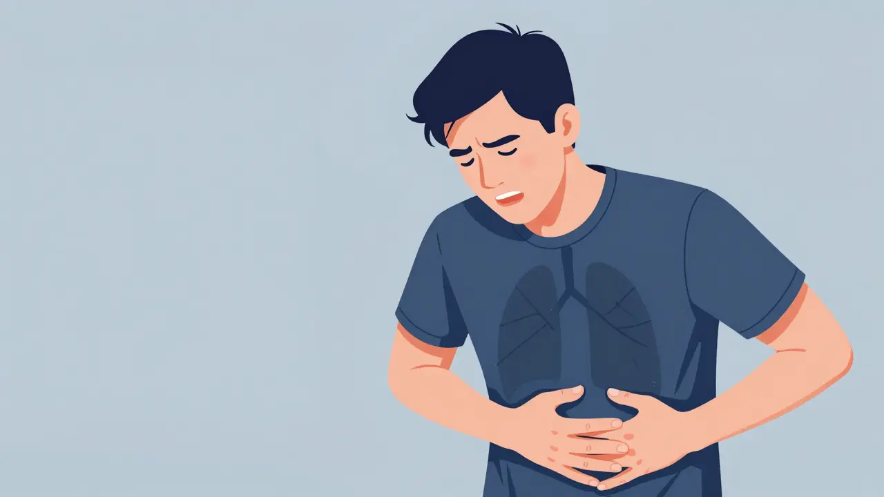
Imagine taking a deep breath, expecting air to fill your lungs, only to feel that rush stop abruptly. That sensation is the hallmark of Pneumothorax, medically known as a collapsed lung. This condition occurs when air leaks into the space between your lung and chest wall-the pleural space. Instead of expanding freely, the lung collapses under pressure. According to recent data, this affects approximately 7.4 to 18 per 100,000 people annually, making it far more common than many realize.
The condition was formally described in medical literature back in 1819 by René Laennec, the inventor of the stethoscope. While we have modern technology today, the core clinical value remains unchanged: rapid recognition saves lives. If you experience sudden breathing trouble, treating it as an emergency until proven otherwise is the safest approach.
Recognizing the Warning Signs
Your body usually screams for help when a lung is collapsing, though the volume depends on how severe the air leak is. The most consistent symptom across almost every case study is acute pleuritic chest pain. Think of it as a sharp, stabbing ache localized to one side of your chest. It gets worse if you try to take a deep breath or cough. In nearly 92% of clinical cases, this pain might also radiate toward your shoulder on the same side.
Breathlessness is the other major red flag. Statistics show that between 85% and 92% of patients report shortness of breath. Here is a rule of thumb: if your lung collapse is minor (less than 15%), you might only feel winded during exercise. However, if the collapse exceeds 30%, you will likely feel breathless even when sitting still. Listen to your chest too. A doctor looking for physical exam findings checks for decreased breath sounds on the affected side, which is present in 98.7% of confirmed cases. If you tap on your chest during an exam, it might sound hollow or hyperresonant compared to the healthy side.

The Danger Zone: Tension Pneumothorax
While some cases are manageable with observation, a subset progresses to a life-threatening complication called tension pneumothorax. This is a true medical emergency. In this scenario, air continues to enter the pleural space but cannot escape, building massive pressure that pushes against your heart and other lung.
You need to know the specific measurable parameters here. If a patient’s heart rate spikes above 134 beats per minute, systolic blood pressure drops below 90 mmHg, or oxygen saturation falls below 90% on room air, time is running out. Tracheal deviation-where your windpipe shifts away from the affected side-is a late sign seen in only about 32% of cases according to trauma registries. Do not wait for this sign to act. Guidelines mandate that suspected tension pneumothorax requires needle decompression within 2 minutes of identification, often before confirmatory imaging is even possible.
| Type | Typical Cause | Incidence |
|---|---|---|
| Primary Spontaneous | No underlying disease | Common in tall, thin smokers |
| Secondary Spontaneous | Underlying lung disease (COPD) | Higher mortality risk (16.2%) |
| Traumatic | Injury or penetration | Requires immediate intervention |
How Doctors Diagnose the Leak
When you get to the emergency department, speed matters. Diagnostic imaging is the standard tool to confirm the diagnosis, but the choice of method changes the accuracy. A chest X-ray remains the initial standard with a sensitivity of 85-94% for detecting the condition. However, if you are lying flat-common in trauma situations-its ability to detect the leak drops significantly to 40-70%.
For a clearer picture, CT scans offer near 100% sensitivity and can spot tiny amounts of air, as little as 50mL. Point-of-care ultrasound has become a critical tool for emergency physicians. When performed by experienced hands, the 'lung point' sign provides a specificity of 98.7%. Arterial blood gas analysis helps assess the impact on your body, typically revealing hypoxemia (low oxygen levels) in 78% of cases. The goal is to diagnose quickly because the society of thoracic surgeons reports that diagnosis-to-treatment times average around 22 minutes for stable cases, dropping to just 8 minutes for critical ones.

Treatment Protocols and Recovery
If your pneumothorax is small, you might avoid invasive procedures. For a small primary spontaneous pneumothorax (less than 2 cm rim), observation with supplemental oxygen works well. About 82% of these cases resolve spontaneously within 14 days. Oxygen therapy isn't just about comfort; it accelerates the resorption of air, increasing resolution rates from 1.25% to 4.2% volume reduction per hour.
However, larger cases require active intervention. Needle aspiration has a 65% immediate success rate for primary cases. If that fails or for traumatic cases, chest tube insertion is the go-to treatment. A 28F chest tube achieves a 92% success rate. Surgical options exist for recurrent issues. Video-assisted thoracoscopic surgery (VATS) offers a 95% success rate at one year but involves hospitalization. Preventive measures are crucial because recurrence is real; roughly 15-40% of primary cases come back within two years.
Living With and After Pneumothorax
Once you recover, lifestyle choices play a massive role in preventing a return visit. Smoking is the single biggest risk factor. Quitting reduces the recurrence risk by 77% within one year compared to continuing the habit. You should also respect activity restrictions. The FAA guidelines suggest avoiding air travel for 2-3 weeks after the lung has fully resolved. Scuba diving is generally off-limits indefinitely without surgical intervention due to pressure changes that could cause re-injury.
Follow-up care is non-negotiable. A chest X-ray at 4-6 weeks confirms complete healing. Studies show that 8% of patients develop delayed complications if monitoring stops too early. If you ever feel sudden worsening of pain or new blue discoloration of the skin (cyanosis), call emergency services immediately. These signs appear in 94% of recurrent cases and demand instant attention.
Can a collapsed lung heal on its own?
Yes, small pneumothoraces often heal without surgery. Approximately 82% of small primary spontaneous cases resolve spontaneously within 14 days if treated with observation and supplemental oxygen.
What are the signs of tension pneumothorax?
Warning signs include a heart rate over 134 bpm, blood pressure below 90 mmHg, oxygen saturation below 90%, and tracheal deviation. These indicate a life-threatening emergency requiring immediate decompression.
Does smoking increase the risk of recurrence?
Smoking is a major risk factor with an odds ratio of 22.1 for heavy users. Quitting reduces the risk of recurrence by 77% within one year, making cessation the most critical intervention.
How is pneumothorax diagnosed accurately?
Diagnosis typically starts with a chest X-ray (85-94% sensitivity). CT scans are the gold standard with near 100% sensitivity, and ultrasound is highly effective in emergency settings when done by experts.
When should I seek emergency care?
Seek immediate care if you have sharp chest pain worsened by breathing, sudden shortness of breath, or bluish skin. Severe symptoms like dizziness or rapid heartbeat suggest tension pneumothorax.
Rohan Kumar
March 29, 2026 AT 00:34Another day another reason to believe hospitals run on some secret algorithm for breathing control. 😱 Why does the lung leak air but never the heart? Probably designed that way by the deep state med corps. 💊 We get told to monitor oxygen levels but who monitors the monitor? Seems suspicious honestly. 😒
Sabrina Herciu
March 30, 2026 AT 10:44The statistics regarding spontaneous resolution rates are actually quite encouraging! However!!! Please note that observation periods must be strictly monitored!!! Many patients misinterpret mild symptoms as normal discomfort!! If you experience tachycardia above 134 bpm you MUST seek immediate care!!! This is not something to ignore!!!
walker texaxsranger
March 31, 2026 AT 05:52tension physiology misunderstood often in literature hemodynamic collapse precedes radiographic signs clinical assessment vital CT scans delay treatment window need faster protocols triage protocols inefficient overall mortality risk elevated secondary cases ignored primary focus
Debra Brigman
April 1, 2026 AT 09:51Collapse feels like gravity finally winning the battle against buoyancy inside our chests. A singular tear in the membrane whispers chaos to the rhythm of life. We breathe without thinking until the machine breaks down. Pain becomes a loud teacher demanding our attention to fragility. Existence is just holding pressure against void. It is terrifying to feel the lung give way quietly.
tyler lamarre
April 1, 2026 AT 20:31People exaggerate the severity constantly. Unless you are elite genetic stock the odds are low anyway. Average individuals panic over every breath change. Real professionals know which data points matter and which are noise. Stop treating common conditions like terminal events.
Austin Oguche
April 1, 2026 AT 21:24This information is valuable for public understanding of thoracic emergencies. Cultural differences in symptom reporting often delay diagnosis globally. Patients in high altitude regions may present differently. Clear communication with medical staff remains the priority for recovery success. Thank you for sharing this educational material.
Sarah Klingenberg
April 2, 2026 AT 11:23So nice to see detailed guides like this helping folks :) Knowledge is power especially when health is at stake! Keep taking care of yourselves guys and gals :)
Tony Yorke
April 4, 2026 AT 06:07Stay healthy and listen to your body always.
Rachael Hammond
April 4, 2026 AT 12:46Pain feels different depending on what your body goes through. People forget how fragile the chest cavity really is. It scares me that doctors wait so long sometimes. My uncle had a scare during surgery once. He said the air felt like it was leaking out of him. Breathing becomes such a manual task when things break. You realize how much you rely on automatic functions daily. The stats here are scary if you read past the first paragraph. Smoking kills lungs in slow motion ways mostly. Quitting helps stop the bleeding literally speaking medically. Scuba diving rules make sense after reading this stuff. Pressure changes hurt us even if we dont know why. I hope everyone takes the travel warning seriously for planes. Flying too soon risks popping the seal back up again. Recovery takes patience and listening to your own signs.
Devon Riley
April 5, 2026 AT 12:29Your perspective on recovery is really touching and shows great insight 🤗 Taking care of yourself is the most important thing we can do for our loved ones too ❤️ Wishing everyone smooth healing paths ahead 🌱